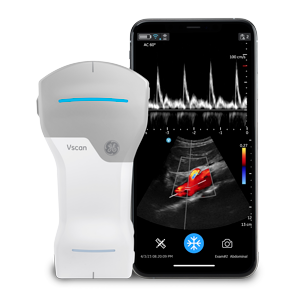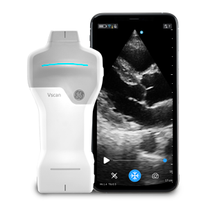
The use of point-of-care ultrasound (POCUS) at the bedside has provided solid clinical benefits, according to the head of a program that has trained internal medicine residents and faculty in its use for more than 10 years.
The device is an important piece of a toolkit that enables clinicians to excel in practicing evidence-based medicine, said David Tierney, MD FACP, internal medicine residency director at Abbott Northwestern Hospital in Minneapolis. The use of POCUS technology is a fundamental component of the program’s ongoing efforts to infuse physicians’ traditional physical exam with more and better information through the use of diagnostic technology.
Dr. Tierney started the Internal Medicine Bedside Ultrasound (IMBUS) Program in 2009, designing the curriculum used to train internists in both inpatient and outpatient settings. At the time he created the program, very few internal medicine residencies were including robust POCUS training in their curriculum. Dr. Tierney designed his program to learn POCUS longitudinally throughout the 3 years of residency.
“This is truly a three-year longitudinal training program that involves specific rotations, and most importantly for all three years, our residents use POCUS as part of their day-to-day patient care,” Dr. Tierney explained. “The tool is equally valuable in both inpatient and outpatient internal medicine settings.”
The Physical Exam and HHUS
Internal medicine specialists place significant value on the physical examination of patients; designed to be thorough and comprehensive, with appropriate attention to detail as clinicians inspect the body and its systems for illness or injury. In its training program, Abbott Northwestern places particular emphasis on evidence-based medicine, integrating the experience of the clinician, patients’ values and the best available scientific information to guide decisions about care.
The use of POCUS as a technology seemed to fit the organization’s goals, as it armed clinicians with more information on which to base care choices.
“To train internists and residents going forward, we really needed a different tool, because we wanted them to be really good at physical diagnosis without requiring 20 years in practice to get there,” he said. “So, this was the right tool at the right time for a place that has always combined leading-edge technology, medical education, and an evidence-based view of the traditional physical exam.”
That’s not to say that this change in diagnostic approach was easy, he recalled.
“The traditional physical exam may be more important to internal medicine than any other specialty,” Dr. Tierney noted. “It’s part of the foundation of internal medicine, so any time you’re going to attempt to change a pillar in medicine, especially the traditional physical exam– you know you’re going to have resistance.”
The program did end up modifying the traditional exam with POCUS, but practitioners now see the value that it brings to diagnostic efforts.
“There was a lot of work over the years to make clear that there’s a role” for both the physical exam and this technology, Dr. Tierney said. “And when the two come together in a collaborative way, from a physician standpoint, you realize huge benefits in quality of care, safety, patient satisfaction, and efficiency of care delivery. It took a little while to demonstrate that, and it took a lot of conversations.”
Storing POCUS Images in the Cloud
The training program pairs the use of POCUS with a secure cloud-based medical imaging platform that stores ultrasound images and makes it easier to share them. A detailed indexing database enables quick retrieval of images for comparative analysis and training purposes. It serves as a non-hospital PACs solution that addresses the organization’s academic needs from an educational archive standpoint. The Abbott Northwestern archive contains some 2,200 different educational POCUS studies that helps internists train in this new modality.
The cloud-based archive also enables residents to share POCUS images remotely with residency faculty and subspecialists, enabling consultations to occur at any time and from anywhere with an Internet connection. The system improves efficiencies by enabling clinicians-in-training to quickly acquire diagnostic information at the bedside, sometimes avoiding the need to schedule a later, separate imaging study which could delay the delivery of care.
The ability to quickly acquire an image from a POCUS device is especially important in treating patients during the COVID-19 pandemic, Dr. Tierney said. “In a world of COVID, we try to minimize the number of people who enter a patient room – that means not just physicians and nurses, but also ultrasonographers and EKG techs – that’s a role that point-of-care ultrasound has taken. Our residents and faculty often gather point-of-care images of the heart, for example, instead of having echocardiography come in and spend 40 minutes with a patient. Using POCUS is a really effective triage mechanism.”
Looking Forward: Use of AI and Machine Learning
The integration of POCUS with artificial intelligence (AI) and machine learning has the ability to even further improve accuracy and speed of diagnosis at the patient’s bedside amongst a wide spectrum of users.
For example, from an AI standpoint using vast stores of POCUS images may help in identifying concerning images of patients down the road. You can add a little bit of machine learning and AI based on thousands and thousands of images that have been accumulated on patients. So, in the future, if you combine the test results with imaging, and you combine the imaging with AI and machine learning, you have the real potential to achieve more powerful insights for improved patient care.”
A repository of POCUS images will be crucial in enabling this diagnostic progress, he believes. “Having real-life images from the emergency room and from the hospital that are analyzed by machine learning and AI programs will be key in physicians making better clinical decisions at the bedside.”
“POCUS and AI can assist clinicians in rural areas to better assess patients without having to refer them for formal studies at larger medical centers miles away in metro areas”, he added. “The ability to be ‘sure enough’ about a diagnosis often takes additional eyes with more experience. AI can provide a clinician with a lot of eyes at the bedside.”
“It’s can be much more efficient if you don’t need to send everyone down to the Twin Cities from the Iron Range in northern Minnesota to get a formal echo knowing that a bunch of (those studies) end up being normal,” he explained. “It’s just better for patients and physicians, from every aspect to be able to have a streamlined and efficient workflow, and AI will tip that to a safe spot where diagnosis using POCUS will become even more accurate and efficient than it is currently. There will always be the need for formal ultrasound and other diagnostic imaging, but this helps make the use of that resource more efficient.”
In conclusion, Dr. Tierney believes that POCUS will give internal medicine specialists valuable insights into their patients, augmenting their hands-on exam while enabling them to have more information with which to make speedy, accurate diagnoses. And the potential benefits of AI and machine learning will only amplify those benefits.