Dyspnea, the sudden and severe shortness of breath, is a frequent presentation in the emergency department and, notably, is one of the most common reasons for hospital visits. There is a broad range of dyspnea causes — including cardiac, pulmonary, metabolic or shock — making it difficult for ED physicians to diagnose and treat the condition.
Why Is Timely Diagnosis Important?
Treating the wrong cause in a patient with dyspnea can result in rapid deterioration. Therefore, it’s crucial to identify the underlying cause and initiate prompt and appropriate treatment. Doing so can be lifesaving for these patients.
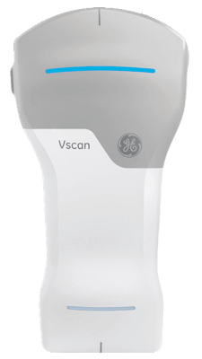
One research paper published in Herz aimed to assess how pocket-sized echo could speed time to diagnosis for patients with acute dyspnea in the ED. It concluded, “The Vscan is a practical, portable device that provides rapid diagnostic information. One-third of patients had significant findings on the scans to possibly aid diagnosis and prevent misdiagnosis.”
Helping Determine the Cause
Traditional tests that include brain natriuretic peptide, chest X-ray, CT pulmonary angiogram or formal echocardiograph can take time to perform and generate results.
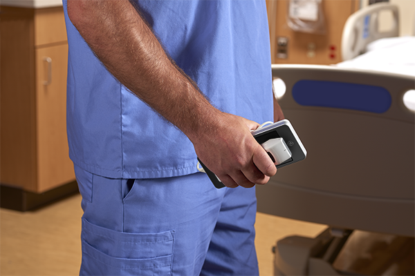
Using handheld ultrasound to diagnose the cause of dyspnea can support a fast diagnosis in an emergency setting and help expedite the implementation of a treatment plan.
One study in the Journal of Hospital Medicine trained internal medicine residents to perform bedside lung ultrasound with a pocket ultrasound. The study concluded, “Lung ultrasound performed by residents with a pocket ultrasound improved the diagnostic accuracy of dyspnea. Two residents undergoing extended training showed a total increase in diagnostic accuracy.”
Differentiating Between Types
There are many potential causes of dyspnea, and it’s essential to differentiate between them to implement early treatment. One of the key diagnostic challenges is to distinguish between cardiac (e.g., acute heart failure) and pulmonary causes. By using a lung-cardiac-inferior vena cava (LCI) handheld ultrasound examination, the accuracy of diagnosis may be improved.
A study in Cardiovascular Ultrasound concluded that “rapid evaluation by LCI integrated ultrasound is extremely accurate for differentiating acute dyspnea due to acute heart failure syndromes (AHFS) from that caused by primary pulmonary disease in the emergency setting.”
This suggests LCI integrated ultrasound may be a useful tool to help improve the speed of diagnosis and, therefore, implement timely decision-making and management plans in the ED.
Helping to Improve Patient Care
Overall, using handheld ultrasound to help diagnose the underlying cause in patients with dyspnea may be an easy skill to learn, quick to use and may support faster treatment decisions compared to other options. This has the potential to result in improved patient care through fast diagnosis and treatment.